
 English
 Español
 Deutsch
 日本語
日本語 Polska
 Français
 한국의
한국의 Українська
 Italiano
 Nederlands
 Türkçe
 Português
 Bahasa Indonesia
 Русский
 中國
中國 हिंदी
हिंदीmitochondria 3D Models
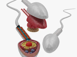
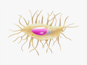
We have 11 item(s) Royalty free mitochondria 3D Models. Buy or download free 3D models for your CG projects, film and video production, animation, visualizations, games, VR/AR, and others. You can download any 3d model in all popular 3d formats including MAX, OBJ, FBX, 3DS, STL, C4D, BLEND, MAYA


Trending searches 3D Models:
Sculpture 3D Models Characters 3D Models Kitchen 3D Models Horse 3D Models Architectural Exteriors 3D Models Phone and Cell Phone 3D Models Vegetable 3D Models Jewellery 3D Models Toys 3D Models Medical 3D Models Helicopter 3D Models Heavy Weapon 3D Models Truck 3D Models Anatomy 3D ModelsQ1: How accurate are mitochondria 3D models for educational use?
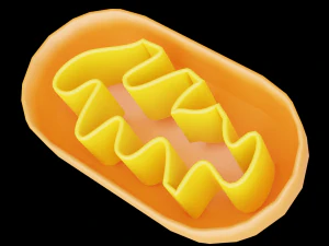
Quality varies considerably. An accurate mitochondria model includes: the outer mitochondrial membrane (smooth ellipsoid or bean-shaped outer boundary), the inner membrane with characteristic cristae folds (shelf-like or tubular invaginations that dramatically increase surface area for ATP synthesis), the intermembrane space between the two membranes, and the matrix (the inner compartment where the citric acid cycle occurs). Cristae geometry is the distinguishing detail — models that show the outer shape without internal cristae miss the most functionally significant structural feature. The best educational models on 3DExport include cross-sectional views or cutaway geometry that exposes the cristae architecture, which is essential for teaching oxidative phosphorylation.
Q2: What educational contexts use mitochondria 3D models most effectively?
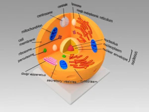
High school and university biology are the primary markets — the mitochondria is covered in virtually every biology curriculum as the site of cellular respiration. The "powerhouse of the cell" meme has made it culturally famous beyond biology classrooms, which drives a wider demand than most organelle models. Medical and pre-med education uses more detailed versions showing the electron transport chain protein complexes embedded in the inner membrane. For interactive educational software, a mitochondria model with separable layers — outer membrane removable to show the cristae, cristae removable to show the matrix — allows progressive reveal teaching that 2D diagrams can't replicate. This layered approach is particularly effective for visual learners.
Q3: Can mitochondria 3D models be 3D printed for classroom use?
Yes — a palm-sized mitochondria model at roughly 10–15cm length prints well in PLA. For a teaching model, the cross-section view is more useful than the whole organelle: cut the model along the long axis, print both halves, and display them open to show the internal structure. The cristae geometry requires enough detail to be recognizable at print scale — cristae folds should have at least 3–4mm depth in the printed model to be visible and handleable. Color differentiation is useful for teaching: outer membrane in one color, inner membrane/cristae in another, matrix fill material in a third. Multi-material FDM printers can do this in one print; single-material printers require separate parts assembled post-print.
Q4: How do I animate ATP synthesis for a biology explainer video using a mitochondria model?
Focus on the ATP synthase (Complex V) — the molecular motor embedded in the inner membrane where ADP + phosphate converts to ATP. Model ATP synthase as a simple turbine-like geometry (the F1 head and Fo base) and animate its rotation — the real enzyme rotates at approximately 100–200 RPM during active synthesis. In Blender, add sphere objects representing ATP molecules being released from the F1 head at each rotation cycle (2 ATPs per rotation on average). Show the proton gradient visually — small sphere particles representing H+ ions flowing through the ATP synthase channel from the intermembrane space to the matrix, driving the rotation. This cause-and-effect animation (proton flow → rotation → ATP release) explains chemiosmosis more effectively than any diagram.