
 English
 Español
 Deutsch
 日本語
日本語 Polska
 Français
 中國
中國 한국의
한국의 Українська
 Italiano
 Nederlands
 Türkçe
 Português
 Bahasa Indonesia
 Русский
 हिंदी
हिंदीright knee joint - female Modelo 3D


BLACK FRIDAY
BIGGEST SALE 70% OFF
$
9.00 USD
- Solicitar suporte ao produto pelo autor

- Formatos disponíveis:
- ID do Item:206261
- Data: 2018-08-11
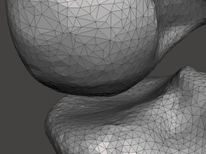
- Polígonos:43,640
- Vértices:21,888
- Animados:No
- Textura:No
- Equipados:No
- Materiais:No
- Low-poly:No
- Coleção:No
- Mapeamento UVW:No
- Plugins Utilizados:No
- Pronto para impressão:No
- Scan 3D:No
- Conteúdo adulto:No
- PBR:No
- Geometria:Polygonal
- UVs não embalados:No
- Visualizações:6289
Descrição
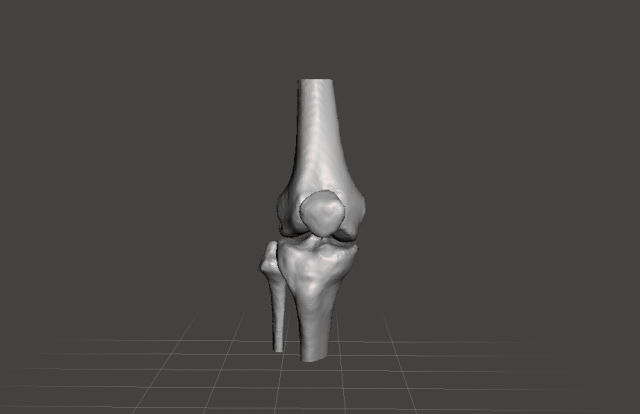
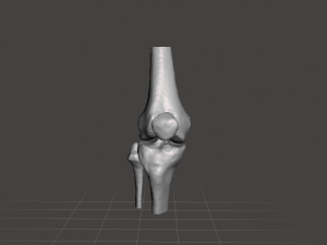
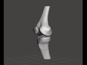
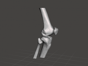
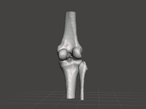
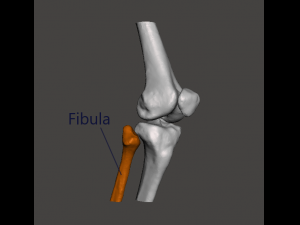
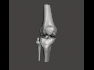
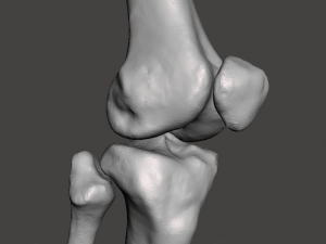
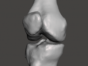
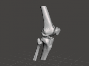
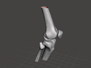
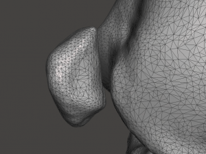
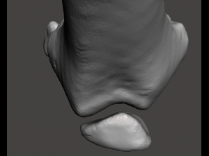
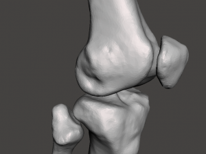
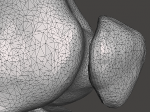
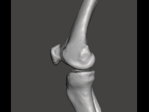
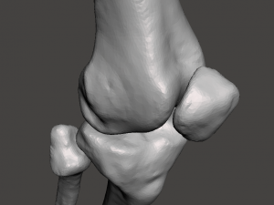
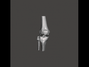
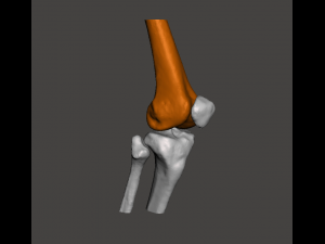
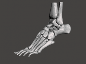
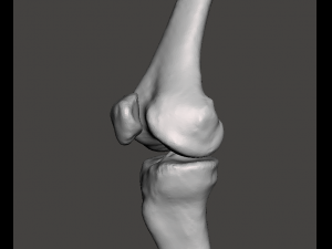
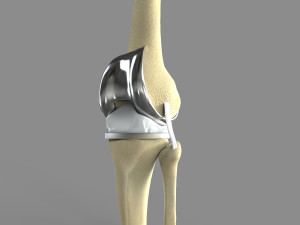
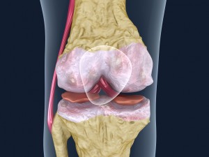
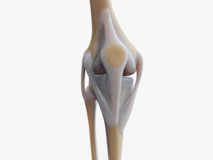
the 3d model was created from computed tomography scans (ct medical data) of the human lower limb. it represents the knee bones (from the right knee).**********
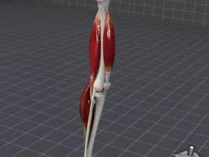
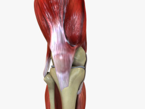
the knee joint is one of the strongest joints in the body. it allows running, walking, standing and supports body weight when it is upright.
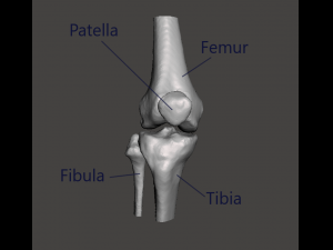
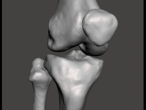
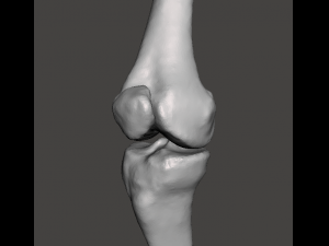
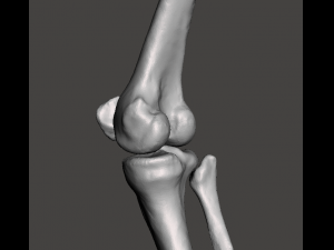
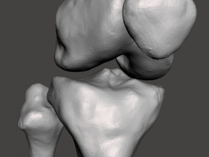
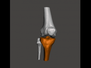
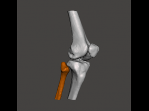
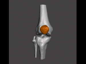
the model includes (from both left side): patella, fibula, tibia and femur
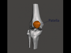
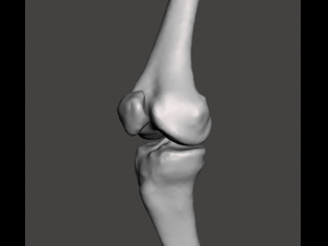
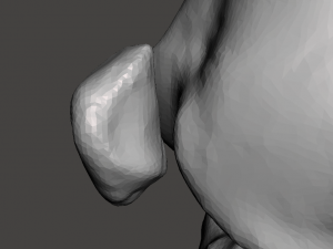
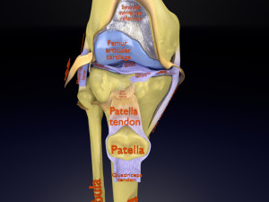
patella, or the kneecap, protects the knee and connects the muscles in front of the thigh to the tibia that play an important role in knee extension.
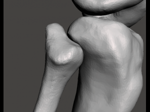
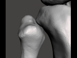
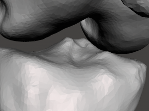
tibia, also called the shankbone or the shinbone, supports most of the weight, and it is the second largest bone in the human body.
fibula, also known as the calf bone, is lateral to the tibia. it stabilizes the ankle. it’s not a weight-bearing bone.
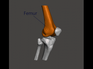
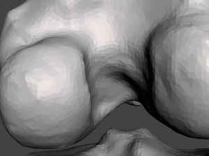
femur (the thighbone) is the longest and the strongest bone of the human body. to break the femur of an average human it would take about 4 000 n. the femur allows motion of the leg. it also supports bodyweight.
patient description: age 45, female
**********
the model was created for educational use.
intends to help those interested in human anatomy and physiology during their learning process, as well as encourage other people deepen their knowledge about the human body. Pronto para impressão: Não
Precisa de mais formatos?
Se precisar de um formato diferente, por favor abra um novo Support Ticket e solicite isso. Podemos converter modelos 3D para: .stl, .c4d, .obj, .fbx, .ma/.mb, .3ds, .3dm, .dxf/.dwg, .max. .blend, .skp, .glb. Conversão de Formato GrátisNão convertemos cenas 3D e formatos como .step, .iges, .stp, .sldprt.!
Informação de utilização
right knee joint - female - Pode utilizar este modelo 3D isento de royalties para fins pessoais e comerciais, de acordo com a Licença Básica ou Prolongada.A Licença Básica abrange a maioria dos casos de utilização padrão, incluindo anúncios digitais, projetos de design e visualização, contas comerciais em redes sociais, aplicações nativas, aplicações web, videojogos e produtos finais físicos ou digitais (gratuitos e vendidos).
A Licença Estendida inclui todos os direitos concedidos ao abrigo da Licença Básica, sem limitações de utilização, e permite que o modelo 3D seja utilizado em projetos comerciais ilimitados ao abrigo dos termos de isenção de royalties.
Leia mais
Vocêm fornecem garantia de devolução do dinheiro?
Sim, fornecemos. Se você comprou um produto e encontrou algum erro nas renderizações ou na descrição, tentaremos corrigir o problema assim que possível. Se não pudermos corrigir o erro, cancelaremos seu pedido e você receberá seu dinheiro de volta em até 24 horas após fazer o download do item. Leia mais condições aquiPalavras-chave
Itens aleatórios do autor
Não há comentários para este item.
-50%
ply stl jpg usd 3mf blend fbx glb glb
deepuparmar777
Anatomy
$149.50
$299.00
-50%
fbx glb blend 3mf usd glb obj stl jpg
deepuparmar777
Anatomy
-50%
3ds c4d fbx lwo lxo ma mb obj stl
clacydarch
Anatomy
$39.50
$79.00
3DDisco
Anatomy
