


 English
 Español
 Deutsch
 日本語
日本語 Polska
 Français
 한국의
한국의 Українська
 Italiano
 Nederlands
 Português
 Bahasa Indonesia
 Русский
 中國
中國 हिंदी
हिंदी
















nefron 3D Modeller
Bizde mevcut 10 ürün(ler) Telifsiz nephron 3D Modeller. CG projeleriniz, film ve video prodüksiyonlarınız, animasyonlarınız, görselleştirmeleriniz, oyunlarınız, VR/AR ve diğerleri için ücretsiz 3D modeller satın alın veya indirin. Herhangi bir 3d modeli, dahil olmak üzere tüm popüler 3d formatlarında indirebilirsiniz. MAX, OBJ, FBX, 3DS, STL, C4D, BLEND, MAYA






Trend olan aramalar 3D Modeller:
Heykel 3D Modeller Karakterler 3D Modeller Mutfak 3D Modeller At 3D Modeller Dış Mimarlık 3D Modeller Ev Telefonu ve Cep Telefonu 3D Modeller Sebze 3D Modeller Mücevher 3D Modeller Oyuncaklar 3D Modeller Tıbbi 3D Modeller Helikopter 3D Modeller Ağır silah 3D Modeller Kamyon 3D Modeller Anatomi 3D ModellerSoru 1: 3DExport'deki nefron 3D modelleri anatomik olarak ne kadar doğrudur?
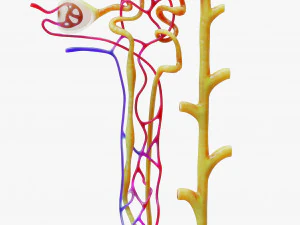
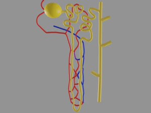
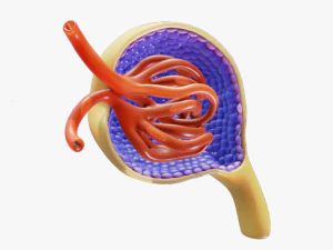
Daha iyi olanlar oldukça detaylıdır — glomerül geometrisiyle Bowman kapsülü, proksimal ve distal kıvrımlı tüpler, hem ince hem de kalın kolları olan Henle kulpu ve toplayıcı kanal. Bu detay seviyesi, nefronu dekoratif bir bilim nesnesi olmaktan ziyade bir öğretim aracı olarak kullanışlı kılan şeydir. Glomerül, yapısal olarak en karmaşık bileşendir — Bowman kapsülü içinde yer alan bir kılcal damar yumağıdır ve bunu basit bir top olarak gösteren modeller, temel fonksiyonel anatomiyi gözden kaçırır. Ciddi eğitim amaçlı kullanım için, tüp segmentlerinin etiketlendiğinden veya renk kodlu olduğundan ve peritübüler kılcal damar ağının, basitleştirilmiş olsa bile, temsil edildiğinden emin olun.
Soru 2: Nefron 3B modelleri hangi eğitim ortamlarında en etkili şekilde kullanılmaktadır?
Üniversite düzeyindeki fizyoloji ve anatomi dersleri birincil pazardır. Nefronun 2 boyutlu diyagramlardan öğretilmesi oldukça zordur çünkü böbrek korteksi ve medullası boyunca izlediği üç boyutlu yol, filtrasyon fonksiyonunu belirler; Henle kıvrımı, idrar konsantrasyonu için gerekli ozmotik gradyanı oluşturmak üzere özellikle medullaya iner. Bu uzamsal ilişkiyi göstermek için döndürülebilen 3 boyutlu bir model, fizyolojiyi anında daha anlaşılır hale getirir. Tıp fakülteleri histoloji laboratuvarlarında etkileşimli nefron modelleri kullanır. Ortaokul biyolojisi, aşırı ayrıntıya girmeden temel filtrasyon-geri emilim-salgılama akışını gösteren basitleştirilmiş versiyonlardan faydalanır.Soru 3: Sınıf içi gösterimler için nefronun 3 boyutlu modeli 3 boyutlu yazıcıda basılabilir mi?
Evet, basılı nefron modelleri dokunsal eğitim yardımcıları olarak iyi sonuç veriyor. Ana tasarım zorluğu tübül geometrisidir: kıvrımlı tübüller dardır (gerçek dokuda 8-60 µm, baskı için elbette büyütülmüştür) ve baskı ölçeğinde kırılgan olabilirler. FDM baskı için, 1:1 eğitim ölçeğinde tübül çapı, elleçlemeye dayanabilmesi için en az 3-4 mm olmalıdır. Reçine baskı daha ince ayrıntıları yakalar ve karmaşık tübül geometrisi için daha iyi bir seçimdir. Renk ayrımı - farklı tübül segmentlerini farklı filament renklerinde yazdırmak - ya çok malzemeli bir yazıcı gerektirir ya da bölümleri ayrı ayrı yazdırıp birleştirmeyi gerektirir. İkincisi, sınıf modelleri için daha yaygın bir yaklaşımdır.Soru 4: Bilimsel yayın için nefron modeli oluşturmak için en iyi yazılım hangisidir?
Yayın kalitesinde render'lar için, Cycles ile Blender ücretsizdir ve KeyShot veya Cinema 4D gibi ticari araçlarla karşılaştırılabilir sonuçlar üretir. Anahtar nokta malzeme kurulumudur: her anatomik bölge, izleyicilerin bileşenler arasındaki mekansal ilişkileri görmelerini sağlayan farklı, yarı saydam bir malzemeye sahip olmalıdır. Tübül duvarlarındaki yüzey altı dağılımı, doku benzeri bir görünüm verir. Dergilere gönderilen şekiller için, en az 300 DPI'da render alın — tipik şekil genişlikleri 8–17 cm olduğunda, bu en az 1000×800 piksel, tercihen 2000×1600 piksel çözünürlük anlamına gelir. İnce anatomik detaylarda sıkıştırma bozulmalarını önlemek için JPEG yerine TIFF veya EPS olarak dışa aktarın.