


 English
 Español
 Deutsch
 日本語
日本語 Polska
 Français
 Українська
 Italiano
 Nederlands
 Türkçe
 Português
 Bahasa Indonesia
 Русский
 中國
中國 हिंदी
हिंदी
















네프론 3D 모델
보유하고 있는 것 9 아이템 로열티 프리 nephron 3D 모델. CG 프로젝트, 영화 및 비디오 제작, 애니메이션, 시각화, 게임, VR/AR 등을 위한 무료 3D 모델을 구입하거나 다운로드하세요. 다음을 포함하여 모든 인기 있는 3D 형식으로 3D 모델을 다운로드할 수 있습니다. MAX, OBJ, FBX, 3DS, STL, C4D, BLEND, MAYA






인기 검색어 3D 모델:
조각 3D 모델 캐릭터 3D 모델 부엌 3D 모델 말 3D 모델 외관 3D 모델 휴대 전화 3D 모델 야채 3D 모델 쥬얼리 3D 모델 장난감 3D 모델 의료 3D 모델 헬리콥터 3D 모델 중무기 3D 모델 트럭 3D 모델 해부학 3D 모델질문1: 3DExport의 신장 3D 모델은 해부학적으로 얼마나 정확한가요?
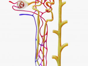
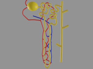
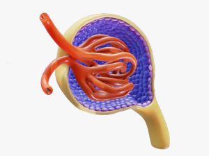
훌륭한 모형들은 상당히 세밀하게 묘사되어 있습니다. 사구체의 기하학적 구조를 보여주는 보우만 주머니, 근위 및 원위 세관, 가는 부분과 굵은 부분을 모두 포함하는 헨레 고리, 그리고 집합관까지 표현되어 있습니다. 이러한 수준의 세밀함 덕분에 네프론 모형은 단순한 장식용 과학 교구가 아닌 유용한 교육 도구로 활용될 수 있습니다. 사구체는 구조적으로 가장 복잡한 구성 요소입니다. 보우만 주머니에 둘러싸인 모세혈관 다발로 이루어져 있는데, 이를 단순한 공 모양으로 표현한 모형은 핵심적인 기능적 해부학적 구조를 놓치고 있습니다. 교육용으로 진지하게 사용하려면 세관 부분에 명칭이나 색깔이 표시되어 있는지, 그리고 세관 주변 모세혈관망이 단순화되었더라도 표현되어 있는지 확인해야 합니다.
질문 2: 어떤 교육 환경에서 신장 3D 모델이 가장 효과적으로 활용되나요?
대학교 수준의 생리학 및 해부학 강좌가 주요 시장입니다. 신장의 피질과 수질을 통과하는 3차원 경로가 여과 기능을 결정하기 때문에 신장 단위(네프론)는 2D 도표만으로는 가르치기가 매우 어렵습니다. 헨레 고리는 소변 농축에 필요한 삼투압 기울기를 형성하기 위해 수질로 내려갑니다. 이러한 공간적 관계를 보여주기 위해 회전 가능한 3D 모델은 생리학적 구조를 즉시 명확하게 이해할 수 있도록 도와줍니다. 의과대학에서는 조직학 실험실에서 상호작용형 네프론 모델을 사용합니다. 중고등학교 생물 수업에서는 과도한 세부 설명 없이 기본적인 여과-재흡수-분비 과정을 보여주는 간소화된 모델을 활용합니다.
질문 3: 교실 시연을 위해 신장 세관 3D 모델을 3D 프린터로 제작할 수 있나요?
네, 3D 프린팅된 신장 세관 모형은 촉각 교육 보조 도구로서 효과적입니다. 주요 설계 과제는 세관의 기하학적 구조입니다. 구불구불한 세관은 실제 조직에서는 8~60µm로 좁고(프린팅을 위해 확대된 크기임) 프린팅 시 파손되기 쉽습니다. FDM 프린팅의 경우, 1:1 교육용 크기에서 세관 직경은 최소 3~4mm 이상이어야 취급 시 손상되지 않습니다. 레진 프린팅은 더 미세한 디테일을 표현할 수 있어 복잡한 세관 기하학적 구조에 더 적합합니다. 색상 분리(서로 다른 색상의 필라멘트로 각 세관 부분을 프린팅하는 것)는 멀티 소재 프린터를 사용하거나 각 부분을 따로 프린팅한 후 조립하는 방식이 필요합니다. 후자의 방식이 교실용 모형 제작에 더 일반적입니다.질문 4: 과학 논문 게재를 위한 신장 단위 모델 렌더링에 가장 적합한 소프트웨어는 무엇입니까?
출판용 고품질 렌더링을 위해서는 Cycles가 포함된 Blender가 무료이며 KeyShot이나 Cinema 4D와 같은 상용 도구와 유사한 결과물을 제공합니다. 핵심은 재질 설정입니다. 각 해부학적 영역에는 구성 요소 간의 공간적 관계를 보여주는 명확하고 반투명한 재질이 있어야 합니다. 세관 벽에 서브서페이스 스캐터링을 적용하면 조직과 같은 질감을 얻을 수 있습니다. 학술지에 제출할 그림의 경우 최소 300 DPI로 렌더링해야 합니다. 일반적인 그림 너비가 8~17cm인 경우 최소 1000×800 픽셀, 가급적 2000×1600 픽셀의 해상도가 필요합니다. 미세한 해부학적 세부 사항의 압축 아티팩트를 방지하기 위해 JPEG보다는 TIFF 또는 EPS 형식으로 내보내십시오.