English
 Español
 Deutsch
 Français
 한국의
한국의 Nederlands
 Türkçe
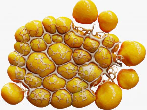
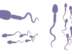
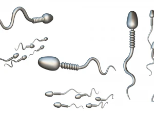
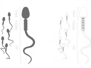
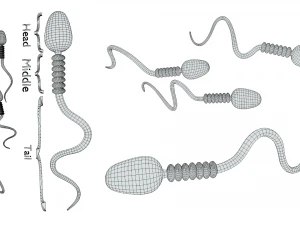
Anatomie der Samenzellen Low-poly 3D Modell

$
9.50 USD
- Fordern Sie Produktunterstützung

- Verfügbare Formate:
- Artikel-ID:468049
- Datum: 2023-10-06
- Polygone:35500
- Eckpunkte:18500
- Animiert:No
- Texturen:Yes
- Rigged:No
- Materialien:Yes
- Low-poly:Yes
- Sammlung:Yes
- UVW mapping:Yes
- Plugins Used:No
- Druckfertige:Yes
- 3D Scan:No
- Erwachsene:No
- PBR:Yes
- Geometrie:Polygonal
- Unwrapped UVs:Non-overlapping
- Betrachter:3081
Beschreibung
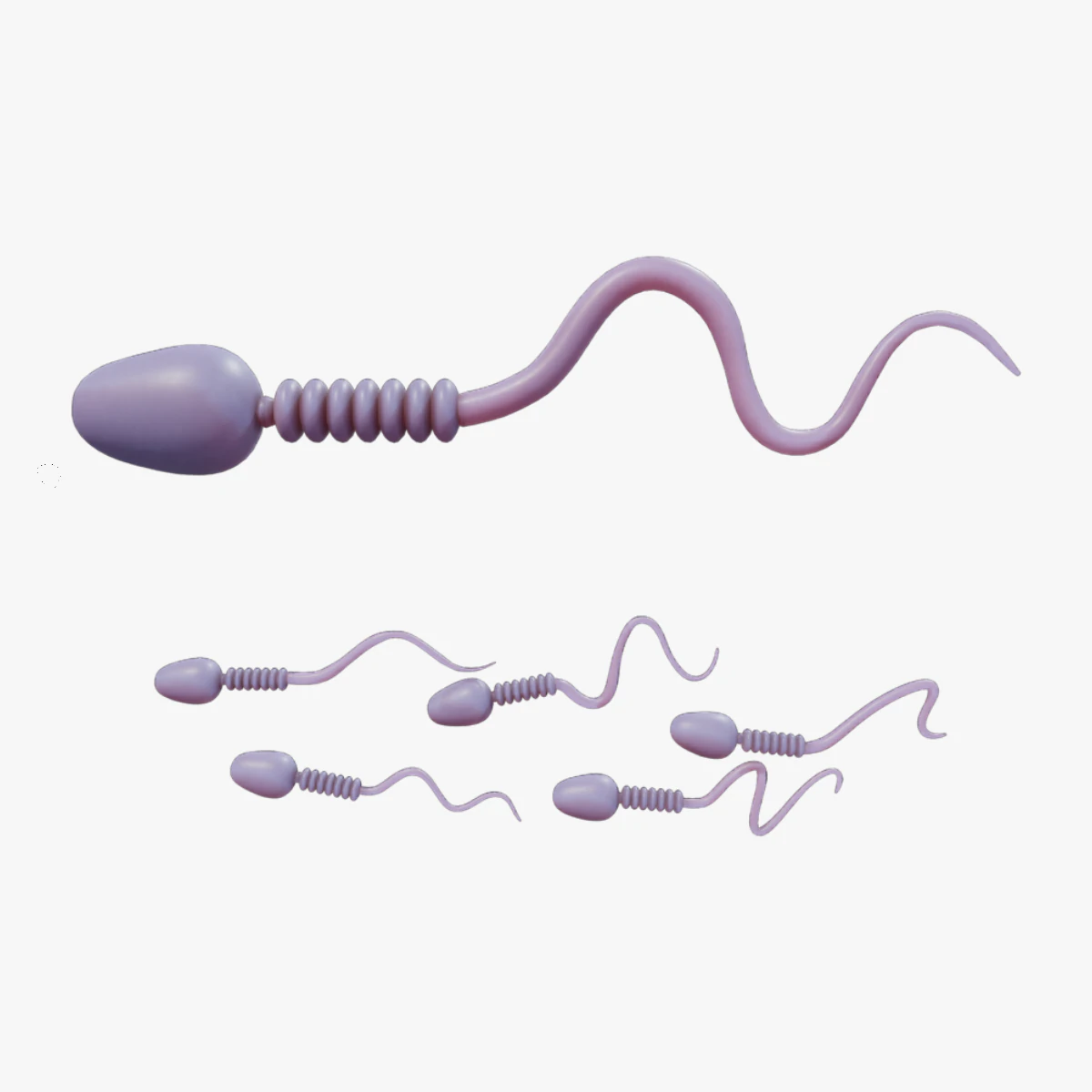
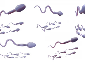
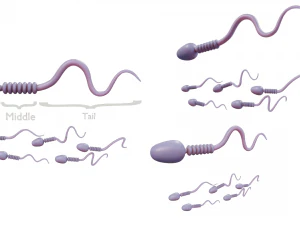
Sperm Cell AnatomySperm develop in the testes and consist of a head, a midpiece, and a tail. The head contains the nucleus with densely coiled chromatin fibers, surrounded anteriorly by an acrosome that contains enzymes for penetrating the female egg.
- Format: FBX, OBJ, MTL, STL, glb, glTF, Blender v3.6.1
- Optimized UVs (Non-Overlapping UVs) (Atlas UV).
- PBR Textures | 1024x1024 - 2048x2048 - 4096x4096 | (1K, 2K, 4K - Jpeg)
- Base Color (Albedo)
- Normal Map
- AO Map
- Metallic Map
- Roughness Map
- Height Map
Sie brauchen mehr Formate?
Falls Sie ein anderes Format benötigen, eröffnen Sie bitte ein neues Support-Ticket und fragen Sie danach. Wir können 3D Modelle in folgende Formate konvertieren: .stl, .c4d, .obj, .fbx, .ma/.mb, .3ds, .3dm, .dxf/.dwg, .max. .blend, .skp, .glb. Kostenlose FormatkonvertierungWir konvertieren keine 3D Szenen und Formate wie .step, .iges, .stp, .sldprt usw!
Nutzungsinformationen
Anatomie der Samenzellen - Sie können dieses lizenzfreie 3D Modell gemäß der Basis- oder erweiterten Lizenz sowohl für private als auch für kommerzielle Zwecke verwenden.Die Basislizenz deckt die meisten Standardanwendungsfälle ab, darunter digitale Werbung, Design- und Visualisierungsprojekte, Social-Media-Konten von Unternehmen, native Apps, Web-Apps, Videospiele sowie physische oder digitale Endprodukte (sowohl kostenlos als auch kostenpflichtig).
Die Erweiterte Lizenz umfasst alle unter der Basislizenz gewährten Rechte ohne Nutzungsbeschränkungen und ermöglicht die Verwendung des 3D Modells in unbegrenzten kommerziellen Projekten unter Lizenzgebührenfreiheit.
Mehr lesen
Bieten Sie eine Geld-zurück-Garantie?
Ja, tun wir. Wenn Sie ein Produkt erworben haben und einen Fehler in den Rendern oder der Beschreibung finden, werden wir versuchen das Problem so bad wie möglich zu beheben. Wenn wir den Fehler nicht beheben können, stornieren wir Ihre Bestellung und Sie bekommen Ihr Geld innerhalb von 24 Stunden nach dem Download des Artikels zurück. . Lesen Sie weitere Bedingungen hierStichworte
Es gibt keine Kommentare zu diesem Artikel.