


 English
 Español
 Deutsch
 日本語
日本語 Polska
 Français
 Українська
 Italiano
 Nederlands
 Türkçe
 Português
 Bahasa Indonesia
 Русский
 中國
中國 हिंदी
हिंदी












마음 3D 모델
보유하고 있는 것 2303 아이템 로열티 프리 heart 3D 모델. CG 프로젝트, 영화 및 비디오 제작, 애니메이션, 시각화, 게임, VR/AR 등을 위한 무료 3D 모델을 구입하거나 다운로드하세요. 다음을 포함하여 모든 인기 있는 3D 형식으로 3D 모델을 다운로드할 수 있습니다. MAX, OBJ, FBX, 3DS, STL, C4D, BLEND, MAYA















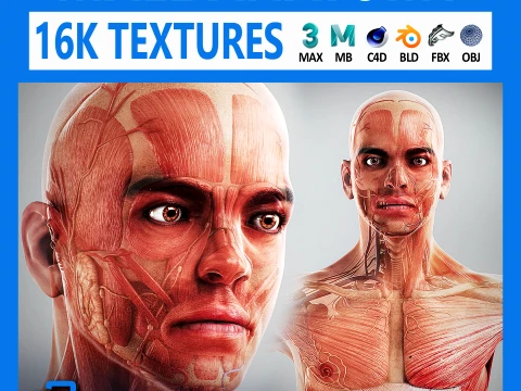













인기 검색어 3D 모델:
조각 3D 모델 캐릭터 3D 모델 부엌 3D 모델 말 3D 모델 외관 3D 모델 휴대 전화 3D 모델 야채 3D 모델 쥬얼리 3D 모델 장난감 3D 모델 의료 3D 모델 헬리콥터 3D 모델 중무기 3D 모델 트럭 3D 모델 해부학 3D 모델Q1: 양식화된 심장 3D 모델과 해부학적 심장 3D 모델의 차이점은 무엇인가요?
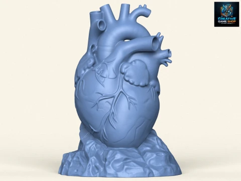
목적과 구조 모두에서 완전히 다릅니다. 발렌타인데이에 흔히 사용되는 하트 모양의 양식화된 하트는 UI 요소, 로고 애니메이션, 장식 소품 등에 유용한 단순한 기하학적 형태입니다. 기하학적 구조는 최소화되어 보통 2,000개 미만의 폴리곤으로 구성됩니다. 반면 해부학적 심장은 완전히 다른 프로젝트입니다. 네 개의 심방(좌심방, 우심실), 대동맥, 폐동맥, 대정맥, 관상동맥 등 각각의 혈관이 정확한 비율로 표현되어야 합니다. MRI 데이터를 기반으로 제작된 해부학적 모델은 실제와 같은 심근 질감을 가지고 있어 수술 시각화나 의학 교육에 활용될 수 있습니다. 이러한 모델들은 별도의 카테고리로 판매되지만, 검색 결과에 함께 나타날 수도 있으니 구매 전에 설명을 꼼꼼히 읽어보시기 바랍니다.질문 2: 해부학적 심장 3D 모델은 의학 교육에 사용할 만큼 정확한가요?
더 나은 모델은 그렇습니다. 참고 자료 출처를 명시한 모델을 찾으세요. 이상적으로는 CT 또는 MRI 스캔 기반이거나 심장 전문의의 검토를 거쳐 모델링된 모델이어야 합니다. 주요 정확도 지표는 다음과 같습니다. 좌심실 벽은 우심실 벽보다 눈에 띄게 두꺼워야 합니다(약 8~12mm 대 3~5mm). 대동맥 판막은 세 개의 첨판을 보여야 하며, 관상동맥은 심외막 표면을 가로지르는 해부학적으로 정확한 경로를 따라야 합니다. 대화형 해부학 소프트웨어 또는 학생 프레젠테이션의 경우, 심실, 판막 및 혈관을 각각 별도의 레이어로 구성한 모델을 사용하면 강사가 구성 요소를 점진적으로 제거하고 표시할 수 있습니다. 이러한 교육 기법은 정적인 다이어그램보다 훨씬 효과적입니다.
질문 3: 의료 훈련용으로 심장 3D 모델을 3D 프린팅할 수 있나요?
네, 3D 프린팅 심장 모형은 수술 계획 및 교육에 사용됩니다. 프린팅을 위해서는 모형이 밀폐되어야 하고 적절한 크기여야 합니다. 성인의 심장은 높이 약 12cm, 너비 약 9cm입니다. 환자 맞춤형 수술 계획을 위해 CT DICOM 데이터를 3D Slicer 또는 Materialise Mimics와 같은 소프트웨어를 사용하여 프린팅 가능한 형상으로 분할한 후, 촉감을 느낄 수 있도록 유연한 TPU 소재로 프린팅합니다. 3DExport의 모형은 환자 맞춤형은 아니지만 해부학 교육용 소품으로 훌륭하게 활용될 수 있습니다. 견고한 전시용 모형은 PLA 소재를 100% 채움 밀도로 프린팅하고, 손으로 만지고 조작할 수 있는 모형은 유연한 필라멘트(TPU 95A)를 사용하세요.
질문 4: 3D 심장 모델을 사용하여 심장 박동을 애니메이션으로 만들려면 어떻게 해야 하나요?
블렌더에서는 셰이프 키와 간단한 리그를 조합하여 사용합니다. 수축기(수축, 부피 감소)와 이완기(팽창, 부피 증가)에 각각 하나씩, 총 두 개의 셰이프 키를 생성합니다. 수축기 셰이프 키의 영향을 심장 주기를 모방한 곡선에 적용하여 애니메이션을 만듭니다. 급격한 수축(0.1~0.15초) 후 느린 이완(0.3~0.4초)이 60~80 BPM의 속도로 이어지도록 합니다. 사실적인 심장 움직임을 표현하기 위해 수축 시 심첨부가 약간 회전하도록(심첨 회전) 미세한 Z축 회전 키프레임을 추가합니다. 모델이 심방과 심실의 형상이 분리되어 있는 경우, 애니메이션 타이밍을 조정할 수 있습니다. 심방이 먼저 수축하고 심실이 그 뒤를 이어 수축하도록 0.1초의 지연 시간을 둡니다.