
 English
 Español
 Deutsch
 日本語
日本語 Polska
 Français
 한국의
한국의 Українська
 Italiano
 Nederlands
 Türkçe
 Português
 Bahasa Indonesia
 Русский
 中國
中國 हिंदी
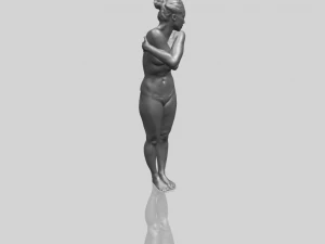
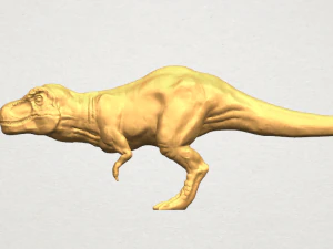
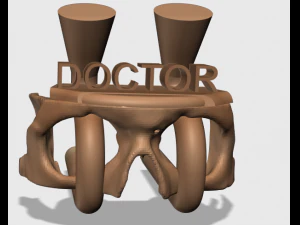
हिंदीBiology - Science 3D Print Models
We have 89 item(s) Royalty free 3D Models. This category contains a wide choice of Biology - 3D Printable Models. Any 3D Printable Biology model is available in .obj, .stl, .iges, .3dm, .skp, .wrl and .blend format. All of these 3d print models are ready for 3D Printing. Also you will find a great number of 3d models in Astronomy and Physics and Engineering categories.
 yassinereyan807Biology
 design3tmBiology


Trending searches 3D Models:
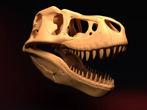
Sculpture 3D Models Characters 3D Models Kitchen 3D Models Horse 3D Models Architectural Exteriors 3D Models Phone and Cell Phone 3D Models Vegetable 3D Models Jewellery 3D Models Toys 3D Models Medical 3D Models Helicopter 3D Models Heavy Weapon 3D Models Truck 3D Models Anatomy 3D ModelsWhat range of biological and medical structures is available for 3D printing?
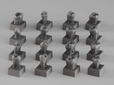
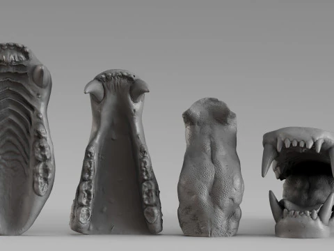
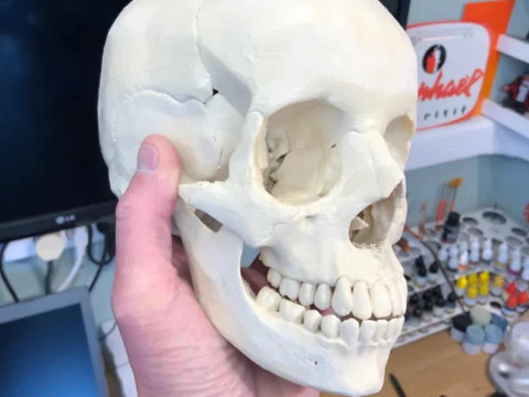
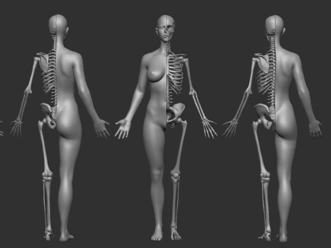
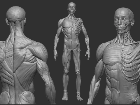
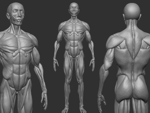
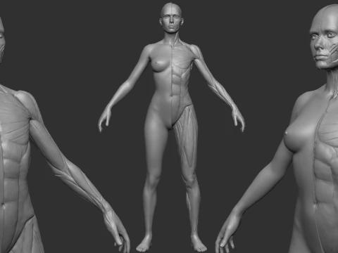
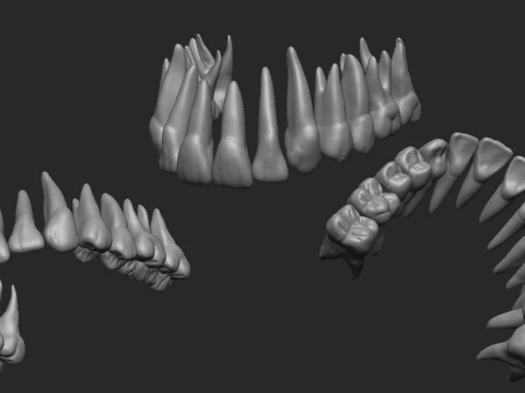
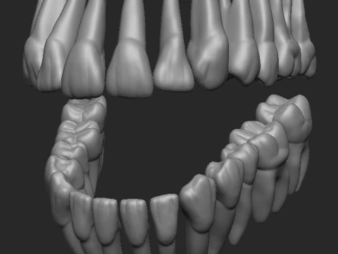
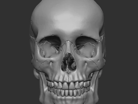
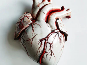
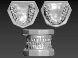
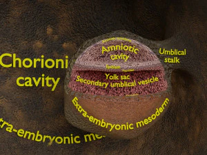
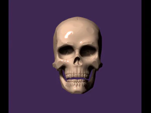
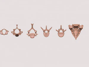
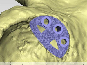
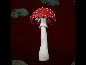
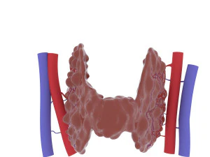
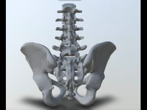
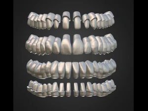
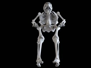
The 2026 Biology category is a comprehensive "Tactile-Life-Library." It covers everything from macro-scale anatomy, such as human skulls and articulating skeletons, down to the microscopic world of cellular organelles, protein folding, and viral capsids. We also provide "Comparative-Anatomy" sets, such as different bird beak shapes or mammalian heart structures. These models are built using "Medical-Scan-Data" (CT and MRI scans) to ensure total anatomical accuracy. They are indispensable for medical students, biology teachers, and museums, providing a physical way to study the complex, organic shapes that define life on Earth, which are often difficult to understand through 2D diagrams alone.
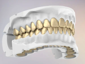
How is the "Internal-Anatomy" made accessible for 3D printing?
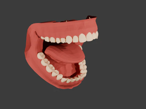
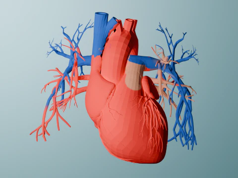
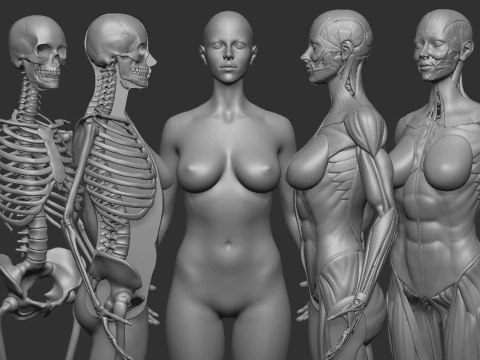
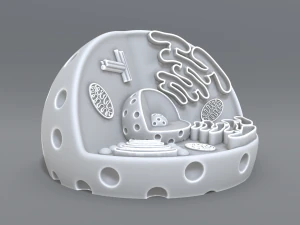
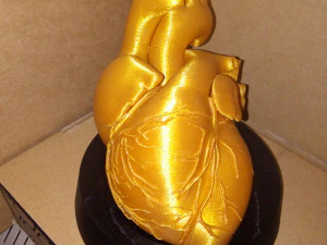
To make internal structures visible, we utilize "Cutaway-and-Exploded" model designs. For example, a 2026 human heart model might be split into magnetic sections, allowing you to "Open" it and inspect the valves and chambers inside. For cellular models, we provide "Nesting-Geometry," where the nucleus and mitochondria are separate prints that fit inside a translucent outer cell wall. This "Modular-Anatomy" approach allows for a "Hands-On" dissection experience that is repeatable and clean. It is a primary requirement for 2026 biological education, as it allows students to physically remove and replace organs or organelles, reinforcing their spatial understanding of how biological systems are organized.
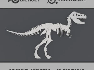
Are the models optimized for printing organic, overhang-heavy shapes?
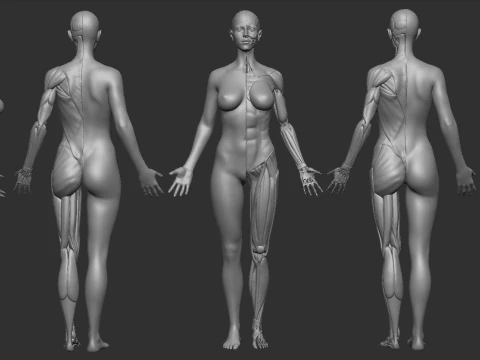
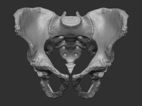
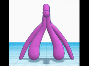
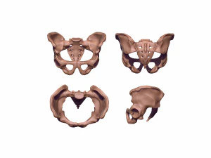
Biological shapes are notoriously difficult to print due to their irregular, organic curves. In 2026, we optimize these meshes by using "Flat-Cut-Planes." For instance, a complex pelvic bone might be provided as two halves that print flat on the bed and are then glued together, which eliminates 90% of the required supports and ensures a beautiful surface finish. For "Full-Body" prints, we provide "Support-Optimized-Stances," where the model is posed to minimize steep overhangs. This technical preparation makes it much easier for educators to print professional-looking anatomical specimens on standard desktop 3D printers without needing expert-level slicing skills.
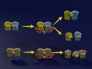
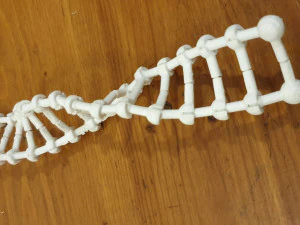
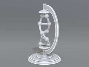
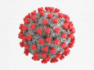
How accurate are the microscopic models of DNA and viruses?
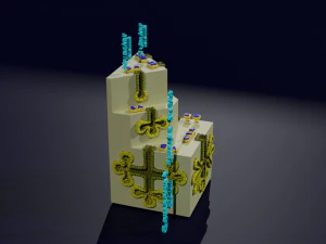
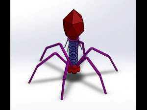
Our 2026 molecular models are based on "PDB-Data" (Protein Data Bank), ensuring that the helical turns of DNA and the geometric symmetry of viral capsids are scientifically precise. These are not "Artist-Impressions," but rather physical visualizations of real molecular structures. In 2026, we also include "Tactile-Binding" features, where different molecules can "Click-Together" based on their actual chemical bonding sites. This makes the models an incredible tool for biochemistry students, allowing them to physically experience how a virus attaches to a cell or how a specific drug molecule fits into a protein receptor, turning "Invisible-Science" into a tangible, interactive learning experience.
What materials and colors work best for biological 3D prints?
For anatomical models, "Bone-White" PLA or matte resins are the industry standard for 2026 as they provide a professional, museum-quality look. However, for "Multisystem" models—like a digestive tract—we recommend using different colored filaments or paints to distinguish between various organs. For cellular models, "Translucent-PETG" is excellent for the outer membrane, while solid colors work for the internal organelles. In 2026, many educators also use "Flexible-TPU" to print lungs or skin sections to give them a realistic, squishy feel. Choosing the right material and color palette is key to making a biological model not just a shape, but a clear and effective communication tool.